Bruker is a performace leader in preclinical imaging instrumentation. Bruker offers nine imaging modalities: PET, SPECT, CT, MRI, MPI, fluorescence, luminiscence, radioisotipic imaging and X-ray.
Discover
Related topics
Extensive assessment of Cytokine production on the NovoCyte Advanteon flow cytometer
Jan 13, 2023
Cytokines are small molecules essential for immune cell response to activation by pathogens, autoimmunity, or...
Microfluidic cell sorter sample preparation for genomic assays
Dec 16, 2022
Single-cell RNA-Sequencing has led to many novel discoveries such as the detection of rare cell populations, microbial...
CellRad+ benchtop X-Ray irradiator
Dec 9, 2022
New CellRad+ benchtop irradiator for biological samples is coming with higher voltage and continue exposure for 180...
Dec 1, 2022
Cytek Cloud is the new digital ecosystem that supports full spectrum flow cytometry research from panel design to data...
See More, Sort More with Cytek Full Spectrum Systems
Nov 28, 2022
Still wondering whether to join the shift to full spectrum cytometry? Easily transfer assays from your Cytek Northern...
Demonstrate the value of RareCyte Orion system via web-based Minerva viewer
Nov 25, 2022
The HTA CRC Atlas X dataset contains images and other data being used for construction of an atlas of human colorectal...
RareCyte Orion system advances Multi-modal digital pathology for colorectal cancer diagnosis
Oct 19, 2022
Precision medicine is critically dependent on better methods for diagnosing and staging disease and predicting drug...
Introducing MARS system : revolutionary solution for your workflow of cell separation and enrichment
Oct 7, 2022
The MARS family of instruments provides a breakthrough solution to complete the workflow of cell separation and...
Automating DNA Extraction for High-Throughput Bacterial Plasmid Purification with CyBi®-SELMA
Sep 19, 2022
The Need to Increase Plasmid Isolation Throughput, Challenges with Scaling up Plasmid DNA Extraction, Automating the...
Introducing RareCyte Orion, integrating Spatial Biology with Multiplex Imaging
Sep 13, 2022
Orion, the highly multiplexed fluorescent imaging system, empower investigators to unlock spatial context for key...
Jan 23, 2017
Historically researchers apply time consuming step serial sectioning and subsequent staining and imaging using light microscopy to analyse the lung morphometry. The data obtained using histology is limited to 2D and, as the pathology is often patchy, the results can be biased and incomplete due to heterogeneous pathological phenotypes. Obtaining an overall status of the entire lung is crucial for reliable and complete data. For instance, when screening the lung for metastasis: these can easily be missed when only a few cross sections are made.
In the method note “MN096_Analysing high resolution lung microCT datasets” a detailed step by step protocol describes how to perform structural analysis of the lung.
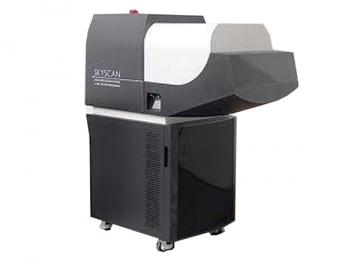
SkyScan software CTAn allows the automatic 3D structure thickness and structure separation analysis from high resolution scans. Structure thickness provides an insight in the distribution of the wall thickness of the airways and / or vasculature of the lungs. In addition, structure separation results in an output of the distribution of the airway diameters, similar as the mean chord length used in histo-pathological analysis. Both types of analysis also allow the possibility to visualize the results in 3D using the SkyScan CTVox volume rendering software.
Related technologies: X-ray and micro computed tomography
Brand profile
Bruker is a performace leader in preclinical imaging instrumentation. Bruker offers nine imaging modalities: PET, SPECT, CT, MRI, MPI, fluorescence, luminiscence, radioisotipic imaging and X-ray.
More info at:
www.bruker.com/