Discover

Back to filter

Related topics

Webinar: Join us for an In-Depth Look at the Revolutionary Small Animal Radiotherapy System at STTARR

Precision X-Ray

Aug 17, 2023

Register for an engaging webinar led by Naz Chaudary, Ph.D., and Research Technician, Alex Wang from the...

Optimizing Gene Expression with Bioluminescence & the piggyBac System

Spectral Instruments Imaging

Aug 15, 2023

Discover how bioluminescence imaging & the piggyBac gene editing system optimize & track gene expression in mouse...

High-plex immunofluorescence imaging and traditional histology of the same tissue section

RareCyte

Aug 7, 2023

RareCyte Orion’ platform has been used for collecting H&E and high-plex immunofluorescence images from the same cells...

Drive key insights and discoveries with NEW Live Cell Analysis applications for eSight system

Agilent technologies

Aug 2, 2023

eSight is now powerful like never before. Learn more about new dedicated application modules for live-cell analysis: 3D...

Single cell-resolution in situ sequencing elucidates spatial dynamics of multiple sclerosis

Vizgen

Jul 14, 2023

MERFISH integrates spatial transcriptomics technology with high resolution spatial imaging, fluidics, image processing,...

High speed, compact and fully automated microscope inside your incubator? Lumascope850

Etaluma

Jul 13, 2023

The powerful, new LS850 Microscope is the latest generation of our fully automated three-channel flagship model and...

MARS - High Efficiency Separation of CD34+ HSC from Mobilized Blood

Applied Cells

Jul 12, 2023

MARS platform provides an easy and cost-effective protocol for CD34+ cell isolation. Single pass CD34+ HSC enrichment...

InAlyzer - The technology chosen by NASA

MEDIKORS

Jul 10, 2023

MEDIKORS's InAlyzer was finally selected as the equipment to be used for NASA's space environment biological research

Nature Article: The genomic landscape of pediatric acute lymphoblastic leukemia

Mission Bio

Jun 29, 2023

Acute lymphoblastic leukemia (ALL) is the most common childhood cancer. Here, using whole-genome, exome and...

Brain-specific biomarkers in urine as a non-invasive approach to monitor neuronal and glial damage

Quanterix

Jun 28, 2023

This study evaluates the quantitative measurability of glial fibrillary acidic protein (GFAP), neurofilament light...

Show all topics (10)

Analysing “tissue sociology” with StrataQuest software

Feb 23, 2016

Cell in tissue plays a defined role as being a member of a small cell subunit within a larger organic structure. The development and differentiation of a given cell is defined by the parent cell and the local cell neighbourhood and environment. Consequently, the analysis of cells in their local environment is of major importance. StrataQuest analysis software contains specific image processing tools for the characterization of the social situation of cells in tissue. Apart from distance measurements there are tools to identify relationships of specific cells by selecting all cells in a local neighbourhood or functional group as a separate population.

The depicted skin sample was stained using the Multi-epitope Ligand Cartography (MELC) method (Fig. A). The tissue sample is subjected to repeated cycles of fluorescent staining, imaging and photo bleaching. Each cycle can use another antibody for a different antigen. The result is a set of images showing the distribution of each antigen in a different layer for the same sample.

The job for StrataQuest in this skin sample was to detect all cells and Melan-A positive areas (Fig. B). Based on this location information, the increase or decrease of the 90 antigen intensities in the areas vicinity was quantified . On this behalf, the Melan-A positive areas were automatically detected and distance transformation masks were established around each of them (Fig.C). The epidermis was also automatically detected (Fig. D).

StrataQuest has the capability to perform these automatic detection, measurement and analysis steps for any functional group of cells in such samples.

Application
Product news

Brand profile

TissueGnostics

TissueGnostics develops systems for automated identification and functional characterization of single cells in tissue sections, cultured and smeared cells as well as TMA. This includes automated image acquisition and image processing for immunohistochemistry and multi-chanel immunofluorescence.

Related products

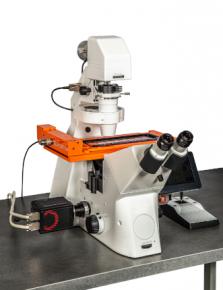
Inverted fluorescence and brightfield system for the scanning and analysis of slides, cytospins, smears and Tissue Microarrays.

show detail

We supply and support Life-Science Technologies in the territory of Central and Eastern Europe.

Czech Republic

Slovakia

Hungary

Poland

Croatia

Slovenia

Serbia

Romania

Bulgaria

Latvia

Lithuania

Estonia

Russia

Ukraine

Belarus

Turkey