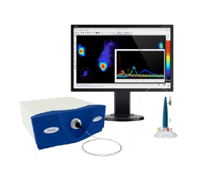
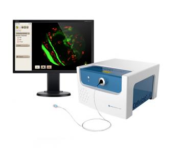
Mauna Kea Technologies manufactures Cellvizio Lab, confocal probe based imaging system, providing cellular-level images with minimal invasiveness for longitudinal studies.
Discover
Related topics
Advances in Leukemia research using shear flow and Bioflux system
Feb 3, 2023
Leukemia is a rare cancer with many subtypes. The production of abnormal leukocytes create disruptions in the immune...
Webinar: Dose Reduction and Image Enhancement in Preclinical Mouse Imaging using Deep Learning
Jan 31, 2023
In this webinar, ir. Florence Muller (Ghent University - University of Pennsylvania) is presenting two recent studies...
Advance your Spatial Biology, customize your panel design with RareCyte Orion reagents
Jan 23, 2023
Orion system, the highly multiplexed fluorescent imaging system, in combination with dedicated reagents, empower...
Extensive assessment of Cytokine production on the NovoCyte Advanteon flow cytometer
Jan 13, 2023
Cytokines are small molecules essential for immune cell response to activation by pathogens, autoimmunity, or...
New installation of Cytek Aurora in Slovenia
Jan 11, 2023
2nd spectral flowcytometer Aurora from Cytek was installed at National Institute of Biology in Slovenia
Microfluidic cell sorter sample preparation for genomic assays
Dec 16, 2022
Single-cell RNA-Sequencing has led to many novel discoveries such as the detection of rare cell populations, microbial...
Dec 1, 2022
Cytek Cloud is the new digital ecosystem that supports full spectrum flow cytometry research from panel design to data...
See More, Sort More with Cytek Full Spectrum Systems
Nov 28, 2022
Still wondering whether to join the shift to full spectrum cytometry? Easily transfer assays from your Cytek Northern...
Demonstrate the value of RareCyte Orion system via web-based Minerva viewer
Nov 25, 2022
The HTA CRC Atlas X dataset contains images and other data being used for construction of an atlas of human colorectal...
Practical aspects of an imaging study: live in vivo scanning
Nov 11, 2022
In this webinar, MOLECUBES will present a live in vivo demonstration, showing the entire workflow from animal...
May 3, 2018
Respiratory diseases, such as pulmonary infections, are an important cause of morbidity and mortality worldwide. Preclinical studies often require invasive techniques to evaluate the extent of infection. Fibered confocal fluorescence microscopy (FCFM) is an emerging optical imaging technique that allows for real-time detection of fluorescently labeled cells within live animals, thereby bridging the gap between in vivo whole-body imaging methods and traditional histological examinations. Previously, the use of FCFM in preclinical lung research was limited to endpoint observations due to the invasive procedures required to access lungs. Here, we introduce a bronchoscopic FCFM approach that enabled in vivo visualization and morphological characterisation of fungal cells within lungs of mice suffering from pulmonary Aspergillus or Cryptococcus infections. The minimally invasive character of this approach allowed longitudinal monitoring of infection in free-breathing animals, thereby providing both visual and quantitative information on infection progression. Both the sensitivity and specificity of this technique were high during advanced stages of infection, allowing clear distinction between infected and non-infected animals. In conclusion, our study demonstrates the potential of this novel bronchoscopic FCFM approach to study pulmonary diseases, which can lead to novel insights in disease pathogenesis by allowing longitudinal in vivo microscopic examinations of the lungs.
Do you want to know more? Read this interesting article!
Brand profile
Mauna Kea Technologies manufactures Cellvizio Lab, confocal probe based imaging system, providing cellular-level images with minimal invasiveness for longitudinal studies.
More info at:
www.cellviziolab.com/