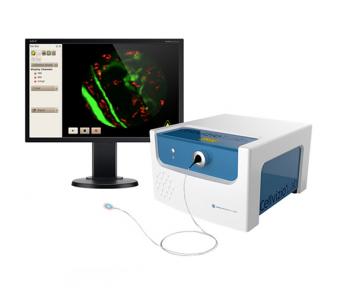
Mauna Kea Technologies manufactures Cellvizio Lab, confocal probe based imaging system, providing cellular-level images with minimal invasiveness for longitudinal studies.
Discover
Related topics
Intoducing spheroOne: Automated sorting and isolation of single spheroids and organoids
Jun 21, 2022
spheroONE is an innovative single large-particle sorter and dispenser which revolutionizes 3D cellular models handling....
Outstanding post-sort sample recovery on the WOLF Cell Sorter
Jun 14, 2022
The new WOLF G2 instrument has significantly expanded the capabilities of gentle benchtop microfluidic cell sorting...
Incucyte AI module: Advanced Label-Free Classification Analysis
Jun 6, 2022
Monitor adherent cell morphology changes objectively to determine live/dead cell counts or classify cells based on...
Favipiravir at high doses has potent antiviral activity in SARS-CoV-2−infected hamsters
Jun 3, 2022
Favipiravir at high doses has potent antiviral activity in SARS-CoV-2−infected hamsters, whereas hydroxychloroquine...
Intoducing cellenONE: The revolutionary Single Cell isolator
May 25, 2022
A revolutionary platform based on sciDROP PICO precision dispensing technology and coupled with advanced image...
Advances in Leukemia research using shear flow and Bioflux system
May 24, 2022
Leukemia is a rare cancer with many subtypes. The production of abnormal leukocytes create disruptions in the immune...
May 4, 2022
A prodigy incorporating a unique combination of patent-pending innovative technologies that takes flow cytometry to the...
In-vivo webinar: a versatile use of SPECT/CT in the development of theranostic tracers
Mar 9, 2022
Combining (preclinical) imaging and targeted therapy, is the basis of theranostics. In this webinar, Dr. Florea will...
Introducing IsoPlexis Single-Cell Natural Killer Panel
Mar 4, 2022
Because of their ability to kill tumour cells, NK cells are an attractive target in cancer immunotherapy, therapeutic...
Enhanced small particle detection on CytekAurora and Northern Lights
Mar 2, 2022
Aurora system with ESP option, can fully resolve particles around 70nm and from the 80nm PS bead. The new violet laser...
Mar 8, 2017
Intraoperative detection of residual malignant cells at tumor margins following excision of primary tumors could help improving surgery and thus patients outcome. The feasibility of the tumor antigens epidermal growth factor receptor and epithelial cell adhesion molecule for antibody-dependent confocal laser scanning endomicroscopy (CLE)-mediated visualization of malignant cells was addressed. Both tumor antigens are highly and frequently expressed in the majority of carcinomas, including head and neck squamous cell carcinomas, and represent prognostic and therapeutic tumor target molecules. FITC-conjugated EGF-R- and EpCAM-specific antibodies served as molecular tools for the detection of antigen-positive cells using the CLE technology. Specificity of both antibodies and their ability to discriminate tumor from non-tumor cells were assessed in vitro with human fibroblasts and PCI-1 HNSCC cell lines, and ex vivo on primary HNSCC samples and healthy mucosa. Antigen specificity of the used EpCAM-specific antibody was superior to that of the EGF-R-specific antibody both in vitro and ex vivo, and allowed visualization of cellular structures in CLE measurements. These results hold promise for possible future applications in humans.
Brand profile
Mauna Kea Technologies manufactures Cellvizio Lab, confocal probe based imaging system, providing cellular-level images with minimal invasiveness for longitudinal studies.
More info at:
www.cellviziolab.com/