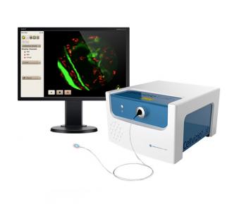
Mauna Kea Technologies manufactures Cellvizio Lab, confocal probe based imaging system, providing cellular-level images with minimal invasiveness for longitudinal studies.
Discover
Related topics
Aug 17, 2023
Register for an engaging webinar led by Naz Chaudary, Ph.D., and Research Technician, Alex Wang from the...
Optimizing Gene Expression with Bioluminescence & the piggyBac System
Aug 15, 2023
Discover how bioluminescence imaging & the piggyBac gene editing system optimize & track gene expression in mouse...
High-plex immunofluorescence imaging and traditional histology of the same tissue section
Aug 7, 2023
RareCyte Orion’ platform has been used for collecting H&E and high-plex immunofluorescence images from the same cells...
Drive key insights and discoveries with NEW Live Cell Analysis applications for eSight system
Aug 2, 2023
eSight is now powerful like never before. Learn more about new dedicated application modules for live-cell analysis: 3D...
Single cell-resolution in situ sequencing elucidates spatial dynamics of multiple sclerosis
Jul 14, 2023
MERFISH integrates spatial transcriptomics technology with high resolution spatial imaging, fluidics, image processing,...
MARS - High Efficiency Separation of CD34+ HSC from Mobilized Blood
Jul 12, 2023
MARS platform provides an easy and cost-effective protocol for CD34+ cell isolation. Single pass CD34+ HSC enrichment...
43 markers, ONE tube : Impress yourself with Cytek Aurora Spectral Cytometer
Jul 11, 2023
Using Cytek full spectrum flow cytometry, scientists at Hamad Medical Corporation, developed a 43 color panel to...
InAlyzer - The technology chosen by NASA
Jul 10, 2023
MEDIKORS's InAlyzer was finally selected as the equipment to be used for NASA's space environment biological research
Nature Article: The genomic landscape of pediatric acute lymphoblastic leukemia
Jun 29, 2023
Acute lymphoblastic leukemia (ALL) is the most common childhood cancer. Here, using whole-genome, exome and...
Brain-specific biomarkers in urine as a non-invasive approach to monitor neuronal and glial damage
Jun 28, 2023
This study evaluates the quantitative measurability of glial fibrillary acidic protein (GFAP), neurofilament light...
Apr 7, 2017
Metastasis of colorectal cancer to the liver is the most common indication for hepatic resection in a western population. Incomplete excision of malignancy due to residual microscopic disease normally results in worse patient outcome. Therefore, a method aiding in the real time discrimination of normal and malignant tissue on a microscopic level would be of benefit.
The ability of fluorescent probe-based confocal laser endomicroscopy to identify normal and malignant liver tissue was evaluated in an orthotopic murine model of colorectal cancer liver metastasis. To maximise information yield, two clinical fluorophores, fluorescein and indocyanine green were injected and imaged in a dual wavelength approach (488 and 660 nm, respectively). Visual tissue characteristics on probe-based confocal laser endomicroscopy examination were compared with histological features. Fluorescence intensity in both tissues was statistically analysed to elucidate if this can be used to differentiate between normal and malignant tissue.
Brand profile
Mauna Kea Technologies manufactures Cellvizio Lab, confocal probe based imaging system, providing cellular-level images with minimal invasiveness for longitudinal studies.
More info at:
www.cellviziolab.com/