When bone experiences large amounts of damage, repair is sometimes beyond the natural ability of the body. Therefore, various techniques have been created over the years to assist in bone reconstruction. In dental surgery, one of the most common to be used is guided bone regeneration (GBR). GBR involves using an absorbable or non-resorbable membrane to shield the wound site from the invasion of soft tissue growth while at the same time applying bone graft material which can provide a scaffold for osteogenic, or bone-producing, cells.
First originating as an experimental procedure in the 1950s, GBR has become well-established in oral and maxillofacial surgery over the last 25 years. However, research continues into how to optimize the technique. For example, comparisons of various membrane types have taken place in animal models. Studies have also looked at how to accelerate bone formation through biological enhancement, such as through the use of growth factors. It has also been explored whether stem cells, taken from the bone marrow, could enhance bone regeneration. While preclinical studies gave positive results, they only used 2D histological analysis, limiting the amount of information they could provide.
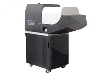
In a recent study, Khalid Al-Hezaimi and colleagues, from King Saud University in Riyadh, Saudi Arabia, showed how 3D micro-computed tomography (CT) could advance preclinical research into GBR.
Read more


