Preclinical in vivo molecular imaging is widely regarded as a key tool within the drug discovery and development pipeline, giving researchers clear visibility of cellular changes at a molecular level. Techniques – or modalities – such as optical, PET and SPECT imaging provide high specificity and wide applicability, as well as the ability to monitor several molecular events and identify key molecular markers. The result is a deeper understanding of disease progression, and the mode of action and pharmacokinetics of potential therapeutics.
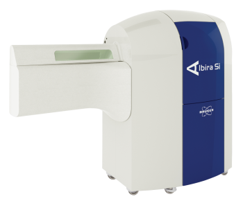
If we consider small animal optical imaging systems specifically, these now enable researchers to combine a number of imaging modalities using the same instrument, thereby paving the way for a better understanding of physiological and disease mechanisms in the preclinical setting. For example, one such system provides five imaging modalities as standard, allowing for co-registration of molecular events with access to bioluminescence, multispectral VIS-NIR fluorescence, unique direct radioisotopic imaging, and Cerenkov radiation. A high speed digital X-ray scanner adds to the functional images with morphological features.
Read more


