Discover

Back to filter

Related topics

The chicken chorioallantoic membrane as a low-cost, high-throughput model for cancer imaging

Precision X-Ray

Apr 4, 2024

Here, we assessed the chicken chorioallantoic membrane (CAM) as an alternative to mice for preclinical cancer imaging...

A microthrombus-driven fixed-point cleaved nanosystem for preventing post-thrombolysis recurrence

IVIM Technology

Apr 3, 2024

A thrombin-responsive and fixed-point cleaved Fu@pep-CLipo was developed for highly efficient and precise thrombolysis...

Multiplexed tissue imaging using the Orion platform to reveal the Spatial Biology of Cancer

RareCyte

Mar 27, 2024

In this webinar Prof. Sandro Santagata, will reveal how Orion high-plex imaging and the use of this data, is valuable...

Innovation is in its DNA

Mission Bio

Mar 22, 2024

Mission Bio’s Tapestri Tool May Hold the Key to Informing Treatment Options for Multiple Myeloma.

A 19-color single-tube Full Spectrum Flow Cytometry for the detection of Acute Myeloid Leukemia

Cytek Biosciences

Mar 13, 2024

This recent publication in Cytometry Part A describes the development and comprehensive workflow of a single-tube,...

18F-labeled somatostatin analogs for somatostatin receptors (SSTRs) targeted PET imaging of NETs

MOLECUBES

Mar 11, 2024

A novel 18F-radiolabeled somatostatin analogue, [Al18F]NODA-MPAA-HTA, was synthesized and evaluated for positron...

Discover Yokogawa CellVoyager CQ1: Benchtop High-Content Analysis System

Yokogawa

Mar 8, 2024

Unlike flow cytometers, the CellVoyager CQ1 confocal quantitative image cytometer does not require pretreatment such as...

Real-time and quantitative analysis of Macrophage Phagocytosis with RTCA eSight

Agilent technologies

Feb 23, 2024

The eSight is currently the only instrument that interrogates cell health and behavior using cellular
impedance
...

Visualization of Tumor-Related Blood Vessels in Small Animals by Photoacoustic 3D Imaging System

Luxonus

Feb 21, 2024

In Vivo Label-Free Observation of Tumor-Related Blood Vessels in Small Animals Using a Newly Designed Photoacoustic 3D...

Therapeutic effects of mesenchymal stem cells in myocardial infarction

S-Sharp

Feb 19, 2024

Intrapericardial hydrogel injection generates high cell retention and augments therapeutic effects of mesenchymal stem...

Show all topics (10)

The most advanced software for preclinical imaging research

Nov 7, 2018

ParaVision is the world‘s leading preclinical imaging software. Used in the most distinguished laboratories, it plays an integral role in the advancement of the cure of diseases, when ground breaking discoveries ranging from basic research to drug development are performed on Bruker instruments run with ParaVision.

Focusing on consistent quantification, ParaVision 360 makes scanning even more intuitive, while maintaining the full flexibility that Bruker users praise. The enlarged MRI sequence portfolio, which is exclusive to Bruker, and the powerful image data evaluation functions lead to maximum proficiency in scanning and analysis.

If you are interesting and if you want to learn more, just read the whole article!

Scientific paper
Application

Related technologies: PET, SPECT, CT

Brand profile

Bruker Biospin

Bruker is a performace leader in preclinical imaging instrumentation. Bruker offers nine imaging modalities: PET, SPECT, CT, MRI, MPI, fluorescence, luminiscence, radioisotipic imaging and X-ray.

Related products

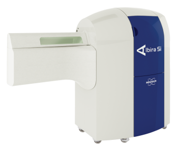
Albira combines PET, SPECT and CT imaging in a unique and extremely powerful way

show detail

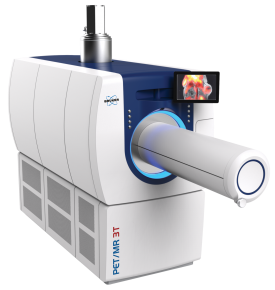
MRI and PET imaging combined for the best of both worlds

show detail

We supply and support Life-Science Technologies in the territory of Central and Eastern Europe.

Czech Republic

Slovakia

Hungary

Poland

Croatia

Slovenia

Serbia

Romania

Bulgaria

Latvia

Lithuania

Estonia

Russia

Ukraine

Belarus

Turkey