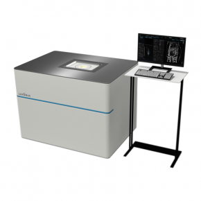
High-contrast images were achieved by increasing the number of integrations during scanning. S-factor images were acquired using both healthy and tumor-bearing mice. Vessels within the liver and kidneys were distinctly reconstructed, and differences in oxygen saturation discriminated between arteries and veins. Repeated measurements on the same mice, both live and post-euthanasia, provided spatio-temporal information, such as a decrease in oxygen saturation after euthanasia or a precipitous drop in oxygen saturation inside the tumor nine days post-cell line transplantation.
Conclusions: By analyzing S-factor images using a photoacoustic imaging system designed for animal experiments, we succeeded in discerning variations in in vivo oxygen saturation. The custom-built system holds promise as a versatile tool for diverse basic research endeavors, as it can seamlessly interface with human clinical applications.
Find entire publication on next link:


